

Klinika dla Małych Zwierząt Wąsiatycz X-ray Unit
-
Take a look at our X-ray facilities!
-
X-ray Unit
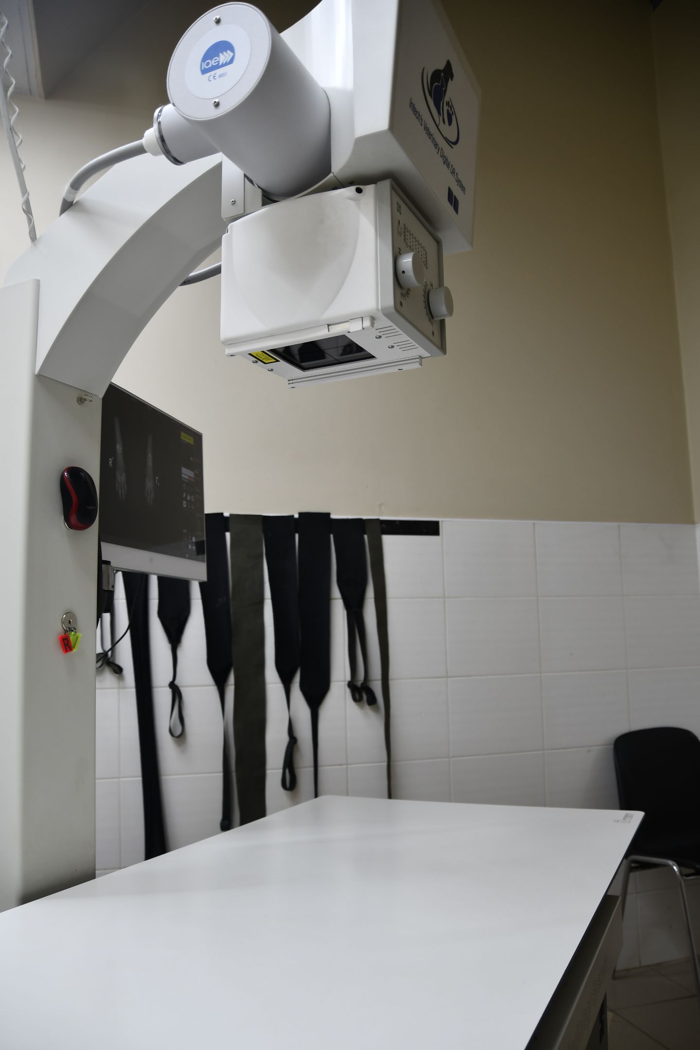
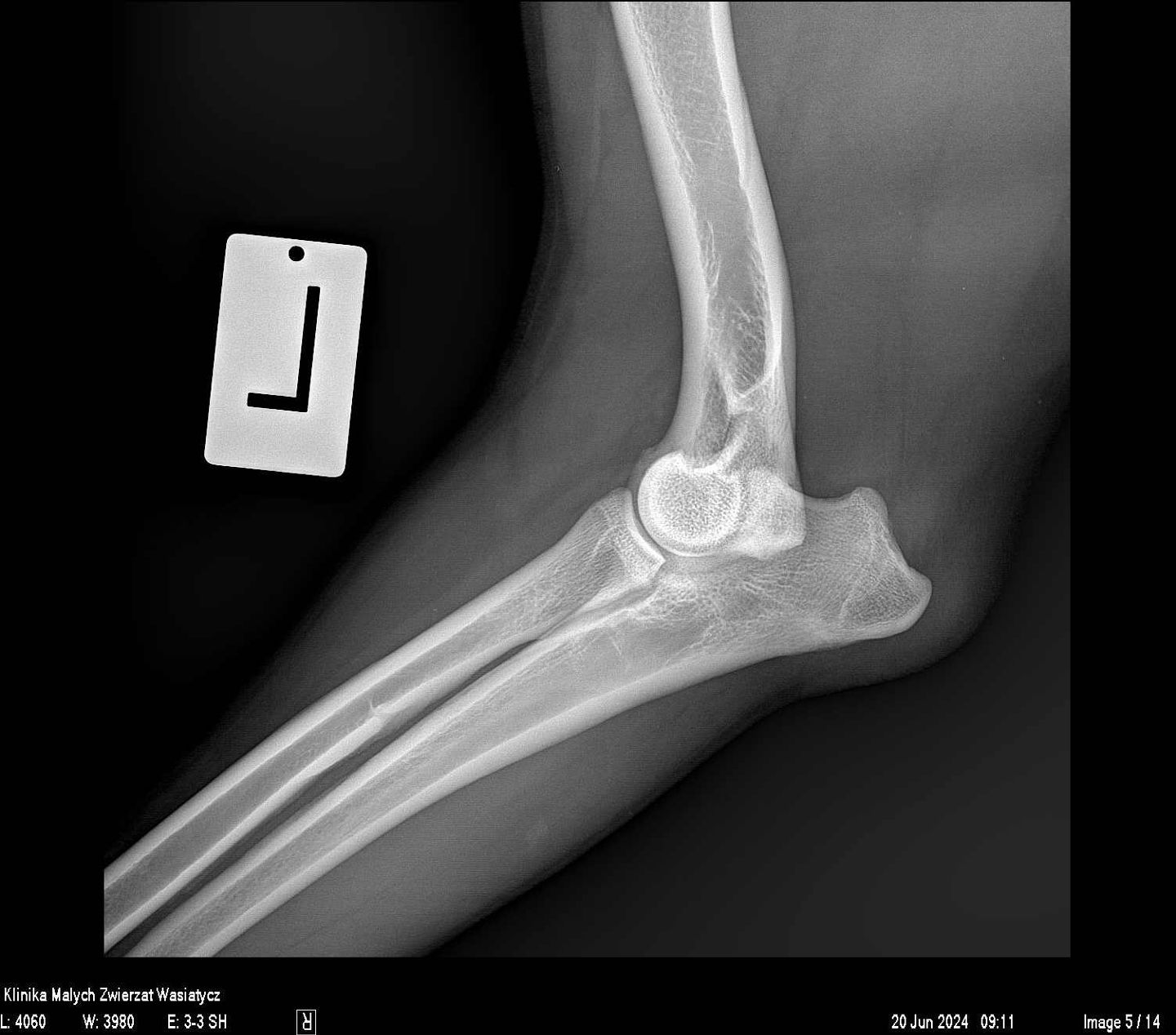
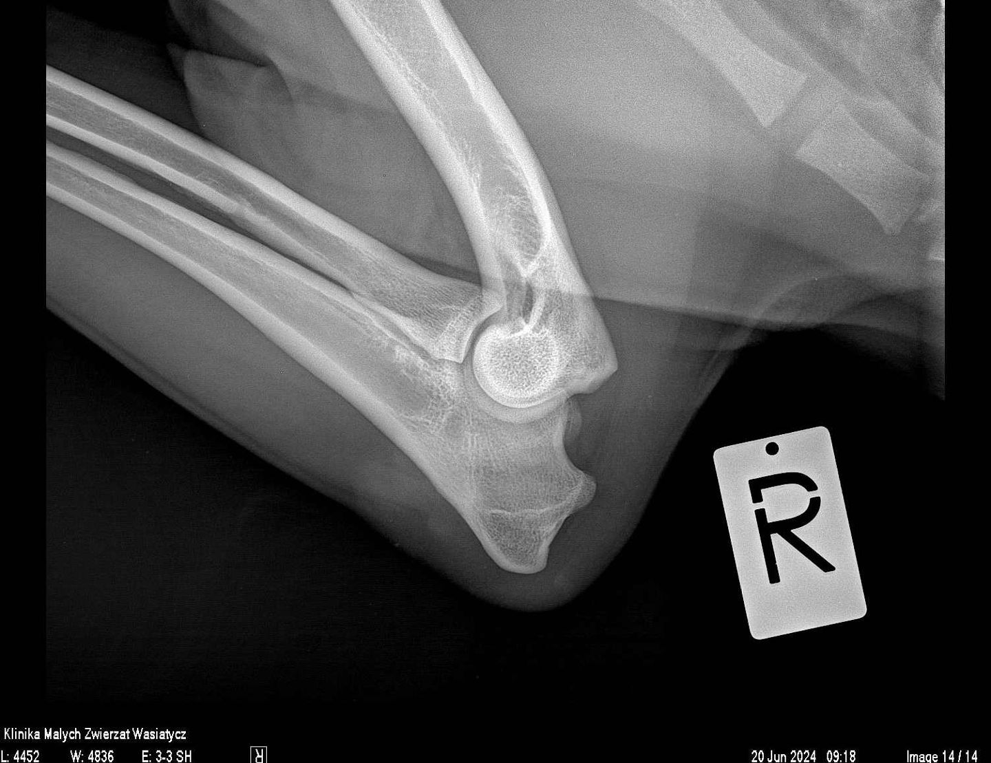
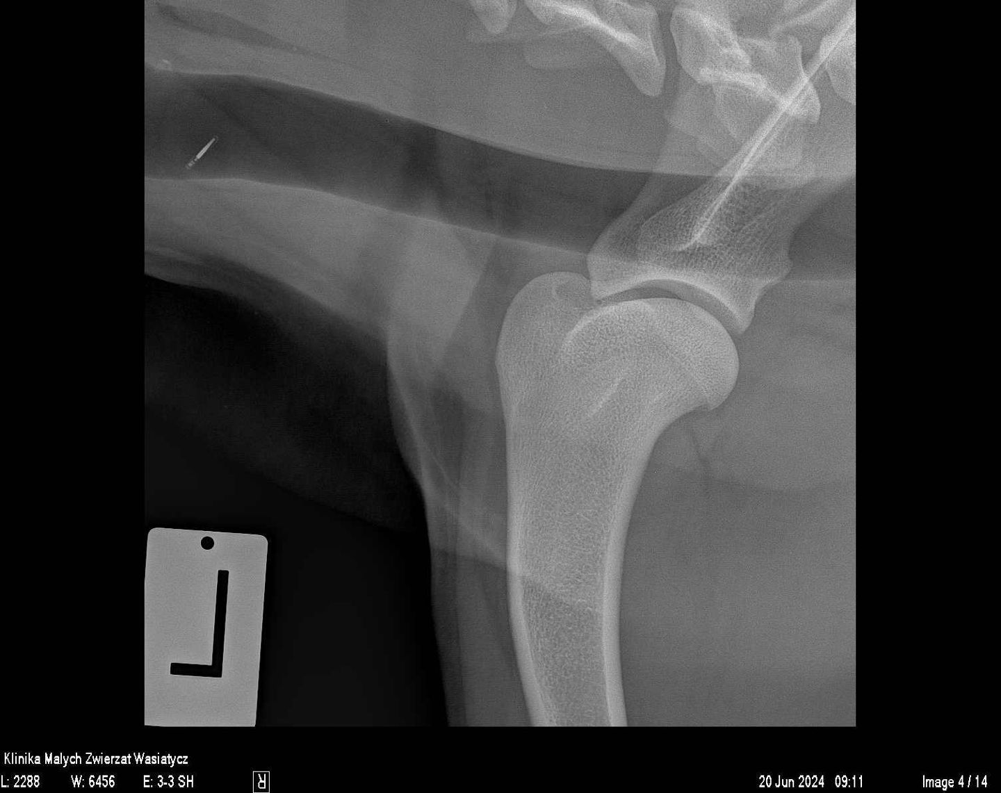
In our clinic, the X-ray Unit has always been a very important diagnostic facility. As a facility specializing in orthopedics, we strive to ensure the highest quality of X-ray images, both in terms of positioning accuracy and exposure parameters.
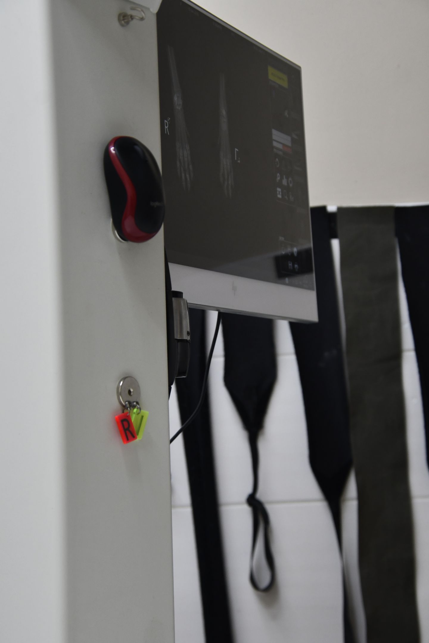
Over 20 years ago, as the first in Polish veterinary medicine, we began using a direct digital radiography system—one where the detector is constantly connected to the computer. This allows for obtaining a finished image just seconds after exposure and sending it to one of the consulting rooms a moment later. At that time, it was one of the world's best X-ray systems from the German company Gierth, based on Canon detectors.
Currently, in our work, we utilize top-class X-ray equipment from the Spanish company Initech, which has been specially configured according to our needs. This is also a direct radiography system.
Examinations using X-rays have been used in medicine for over a century. The development of technology and the use of digital detectors have allowed for a significant reduction in potentially harmful radiation. In current diagnostics, the radiation dose needed to obtain a clear image is up to 50 times lower than in older analogue (film-based) systems.
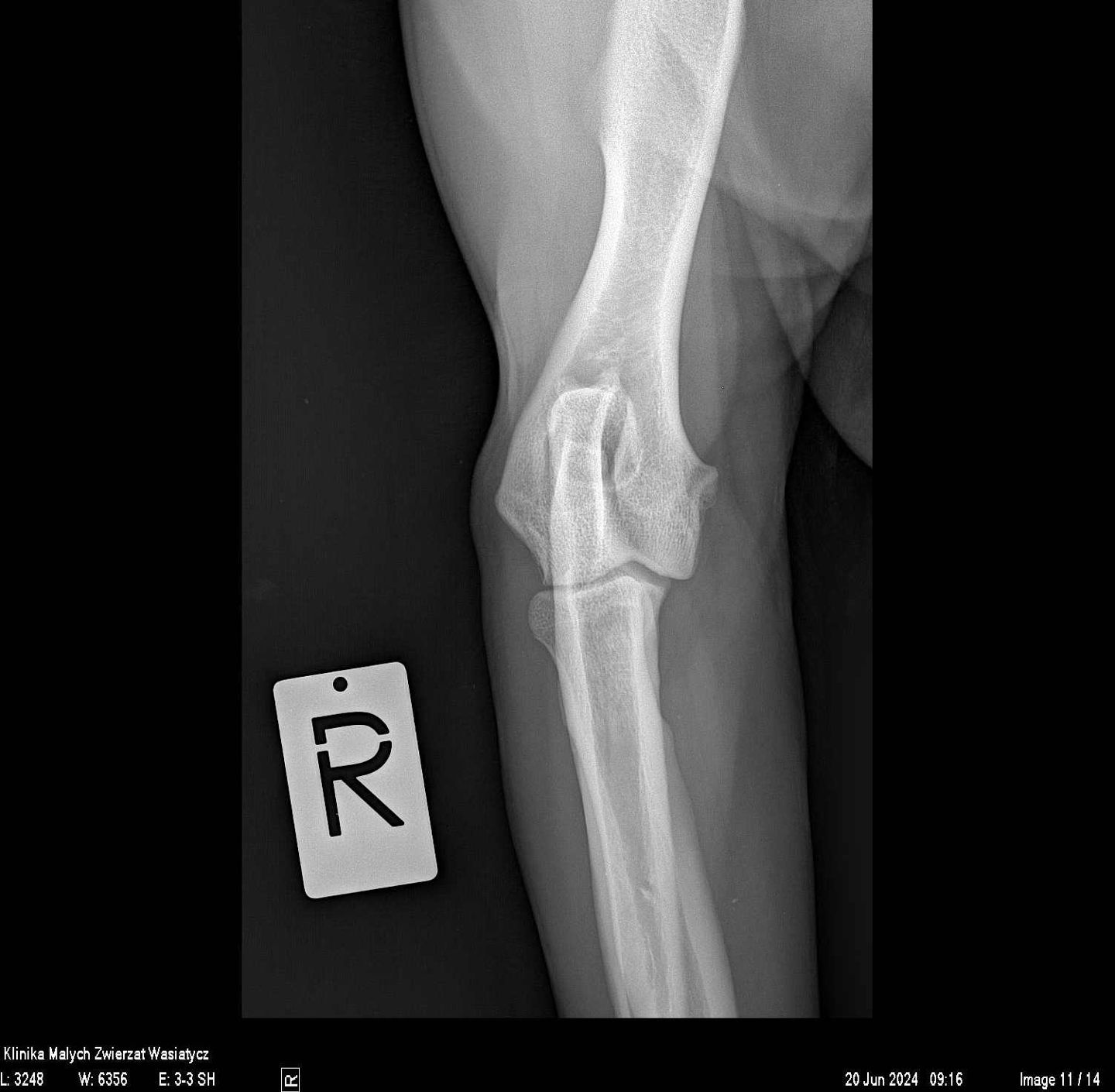
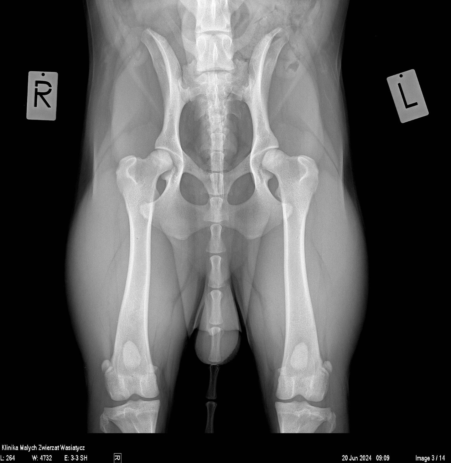
Annually, almost 20,000 X-ray images are taken in our clinic. Each of them is taken by specialists who continuously improve their skills and analyzed by highly qualified veterinarians, including radiology specialists. All of this ensures that the X-ray examination provides the maximum amount of information needed in the diagnostic process.
In our clinic, most X-ray images are taken without sedation of the patient (i.e., without pharmacological calming). Sedation may, however, be necessary in the case of specialised orthopaedic examinations where even muscle tension matters, as well as in situations where diagnostic exposure could cause pain to the animal or if positioning is simply impossible without calming. The animal is held by the owner under the supervision of a trained member of clinic staff, which minimizes the risk associated with administering sedatives and the stress of our patients. All these issues are considered individually and adjusted depending on health status and behavioral matters.
During exposure, the person inside the X-ray room must be equipped with individual radiological shielding (apron, thyroid shield, and if necessary, gloves). It should be emphasized here that the person assisting with the image must not have any contraindications, meaning:
- They must be an adult.
- They cannot be pregnant or breastfeeding.
X-ray images are taken solely on the basis of a PRECISE medical referral, specifying exactly the positioning and the area of examination.
The referral can be:
- Internal, when a doctor during a visit conducted at our clinic needs the aforementioned in the diagnostic process. They then provide exact guidelines to the technician taking the X-rays.
- External (from other doctors, outside our facility) should be in paper or electronic form with the stamp of the facility and/or the referring doctor. It should contain a precise definition of the examination area and the positions needed in the diagnostic process. This is very important because such patients are not seen by a veterinarian but are directed straight to the X-ray Unit.
Images are sent via email to the referring doctor and issued on a DVD to the owner. Due to email transfer limitations, images are sent to the email address (of the referring doctor and/or the owner) as a jpg file. Images recorded on DVD are in the highest quality of medical dicom (dcm) files—they can only be viewed by specialized medical software (one of which is included with the recorded file on the DVD).
NOTE! If the referral does not contain the information necessary to perform the X-ray, a medical visit at our facility will be required to clarify the referral. This involves additional costs in the form of a "first medical visit" fee but is essential for performing images of the greatest diagnostic value and, simultaneously, the lowest possible X-ray dose received by the patient.
It should be emphasized that the people taking images in our clinic are not doctors. Thanks to specialization and continuous improvement in this field, each of the specialists strives for excellence in X-ray work. They are veterinary technicians, an electroradiology technician, and a Master of Medical Physics. Since they are not doctors, they cannot independently decide to change the positioning or the examination area.
After the X-ray images are taken, the finished images are sent to the referring doctor, who is responsible for the description and interpretation of the image. In the case of examinations ordered during a visit, the interpretation of the images occurs a few minutes after the exposure.
Interpretation of X-rays taken on the basis of an external referral belongs to the referring doctor. Obtaining detailed information regarding the images taken and their interpretation is possible only during a follow-up visit at the referring facility.
Contact us
The clinic is open 24/7
Phone Numbers: 61 823 09 97, 61 828 56 01, SMS 799 448 116
e-mail: lp.tewnilkajcpecer
Veterinary Cardiologist - Poznań | Veterinary Ophthalmologist - Poznań | Veterinary Dermatologist - Poznań | Veterinary Neurologist - Poznań | Veterinary Surgeon - Poznań | Veterinary Hematologist - Poznań | Veterinary Endocrinologist - Poznań | Veterinary Orthopedic Surgeon - Poznań | Veterinary Gastroenterologist - Poznań | Veterinary Dentist - Poznań | Veterinary Geriatrician - Poznań | Veterinary Urologist & Nephrologist - Poznań
 This site uses cookies for functional and statistical purposes