

Klinika dla Małych Zwierząt Wąsiatycz Computed Tomography (CT Scan)
-
We were the first in Poland to introduce Cone Beam Computed Tomography (CBCT).
-
How to prepare for a CT scan?
GENERAL RECOMMENDATIONS
- Before the scheduled examination, the patient should fast (no food):
- Ferrets: 5–8 hours,
- Cats and small dogs: 8–12 hours,
- Large dogs: 15–24 hours,
*Rodents and rabbits do not require fasting. - Animals regularly taking medication should receive it on the day of the examination at the usual time.
WHAT TO BRING TO THE EXAMINATION
- The original medical referral for the examination, legibly filled out by a doctor, with a signature and stamp, indicating the location of the lesion/suspected site.
- A complete set of existing medical documentation with previous test results, including: CDs, diagnostic imaging films, discharge summaries from previous treatments, and a list of medications to which the animal is allergic.
- The animal should have laboratory tests performed (results from the last 2 weeks before the CT scan): complete blood count, biochemistry – ALT, urea, creatinine. For older animals or breeds at risk of heart disease, a cardiac examination is also recommended.
- In case of doubts regarding the safety of anaesthesia, you can schedule an anaesthesiology consultation. During such a visit, Wioletta Deoniziak, DVM, will consult on the diagnostic tests performed so far and, if necessary, recommend additional ones. This will allow for a very precise selection of "anaesthetic drugs" and minimize the risks associated with general anaesthesia.
COURSE OF THE EXAMINATION
The Examination:
- Most examinations using computed tomography follow a similar process.
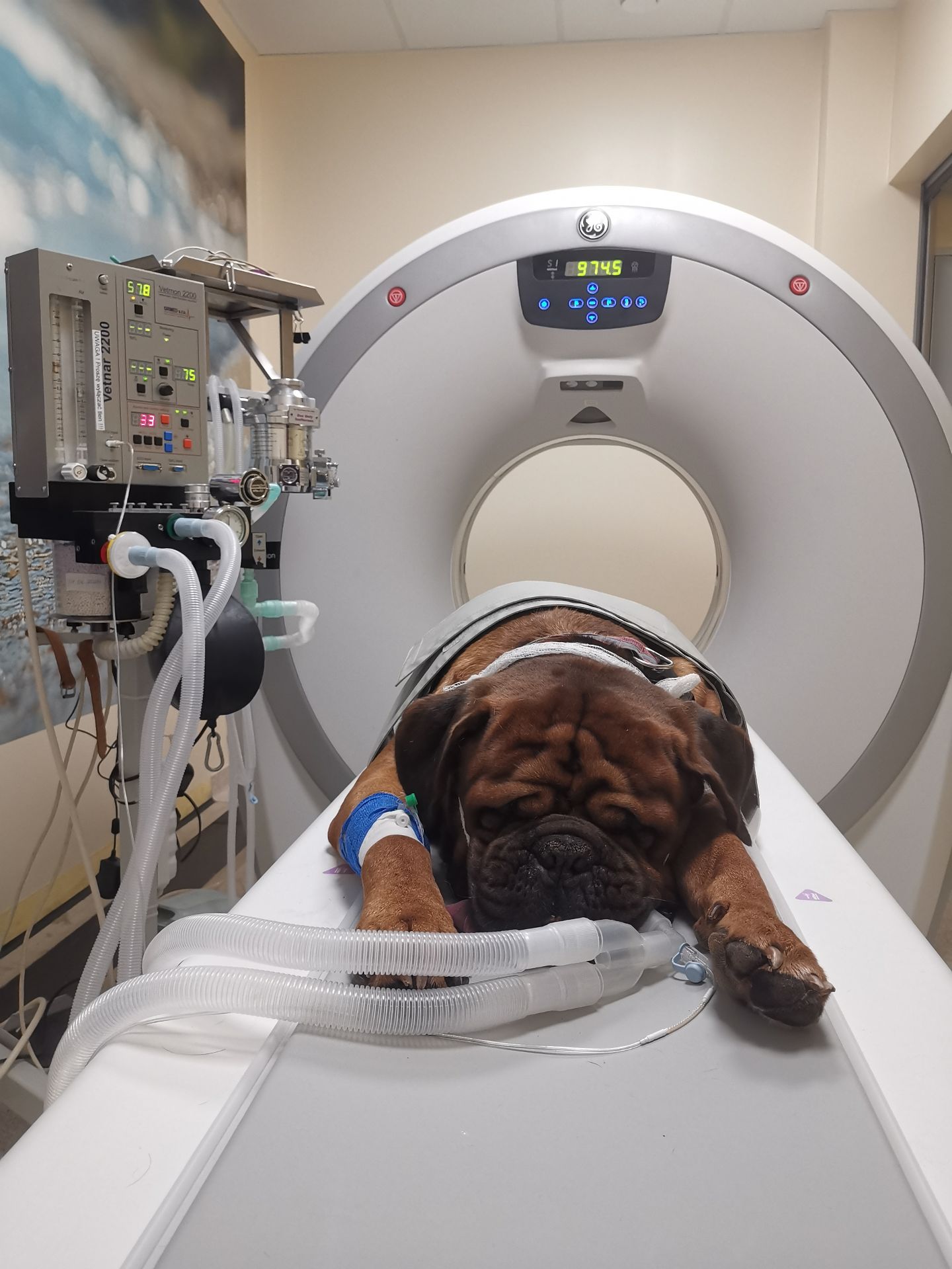
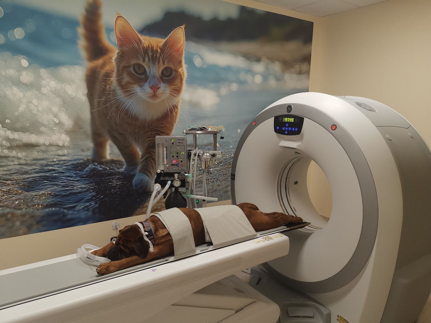
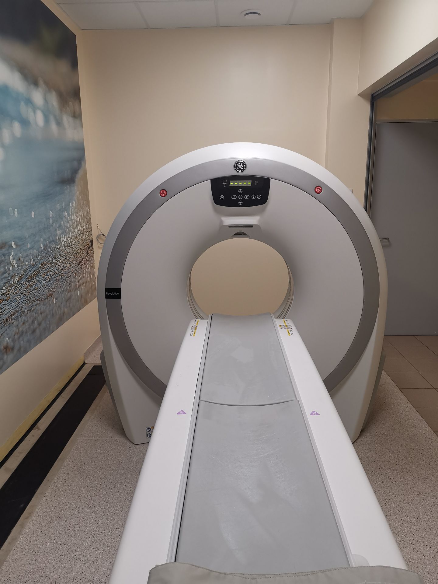
The patient is placed on a special moving table and lies still during the examination while the table moves inside the tomograph. - The medical staff leaves the room containing the CT scanner because the computer station that processes the image information is located in a separate room, from which the technician operates the scanner and monitors the examination.
- The examination is painless. An assistant and a veterinarian watch over the patient.
- In most CT scans, a contrast medium is administered intravenously to obtain the most accurate image of the examined area possible. The contrast medium we apply is very safe and rarely causes allergic reactions.
After the Examination:
- After the examination, the patient remains at the Clinic until it is fully awake. If no adverse effects occur (e.g., contrast allergy or agitation after myelography), only then can the patient leave the facility. After intubation, a cough may appear, which should resolve spontaneously within 24 hours. Nausea and vomiting can be side effects of anaesthesia (even shallow anaesthesia).
- After a spinal examination with subarachnoid contrast administration (myelography), neurological agitation and seizures may sporadically occur, which usually resolve spontaneously within a few to several hours. Until the agitation subsides, the animal must remain under the constant supervision of a veterinarian.
- In the first hours after the examination, do not feed the animal or give it water, and limit physical activity.
Sedation and general anaesthesia can pose a threat to the health and life of the animal. If there are any doubts regarding the patient's behavior after the examination, please contact our Clinic or the nearest open veterinary facility urgently.
Results:
The images obtained during the tomographic examination must be reviewed by a radiologist. This means that you should expect the results within 14 working days. If the "cito" (urgent) option is selected – 3 working days. With the finished description, you should visit a specialist who will use it in the further treatment process.
In August 2014, we launched a method of diagnostic imaging for animals—patients using the computed tomography technique in our Clinic. Until recently, this was the "Fidex" Tomograph, entirely designed and manufactured in the USA.
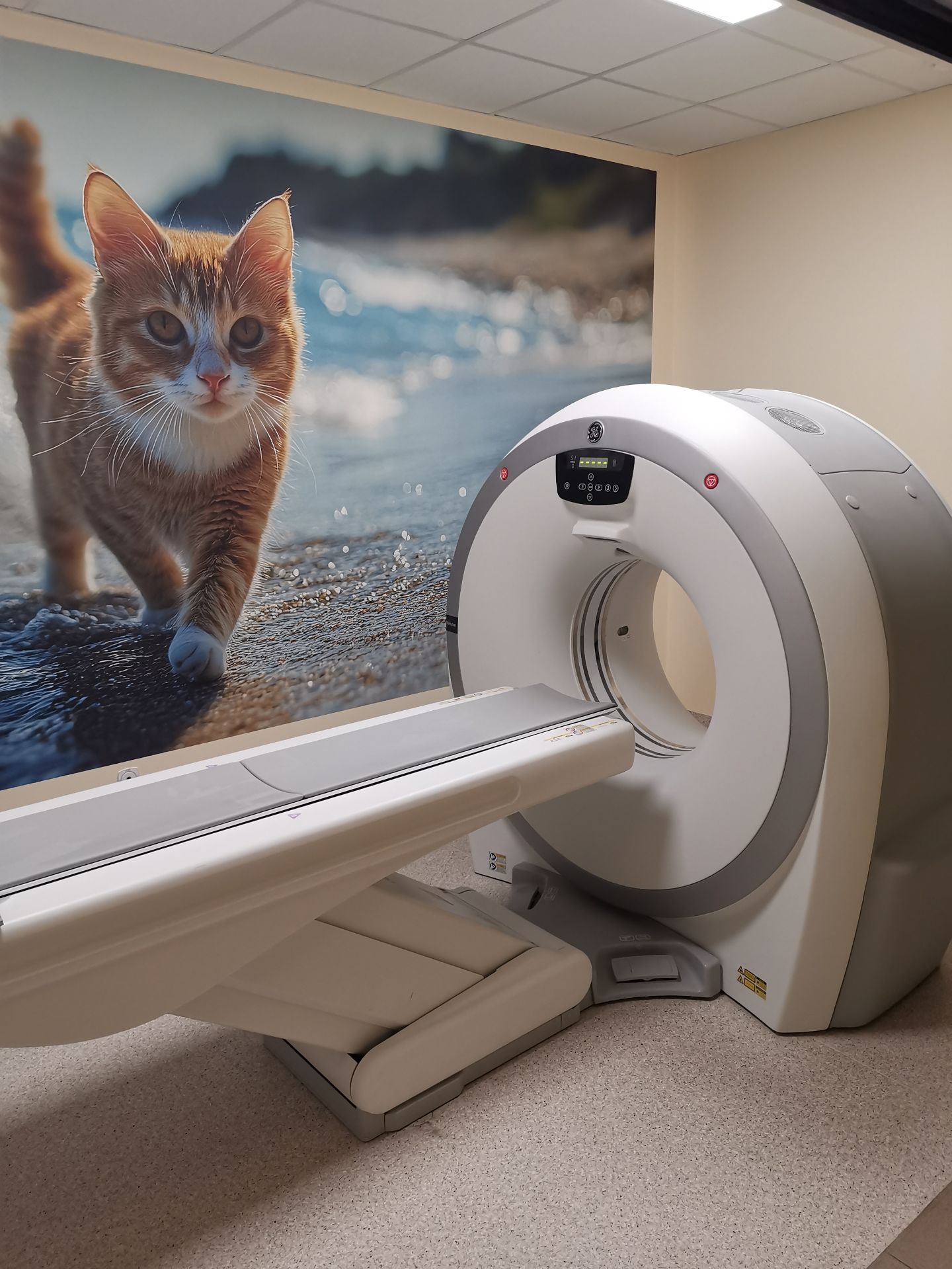
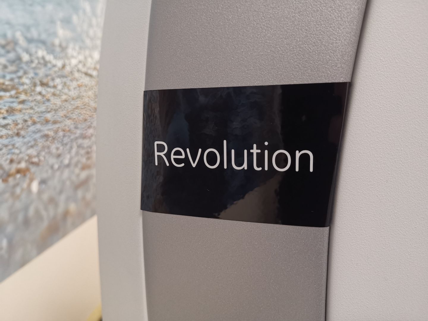
Wanting to increase our diagnostic capabilities, we changed the equipment and currently use a modern 16-slice (16-row) machine from the global manufacturer General Electric. The Revolution ACT 16 LMB computed tomograph is a machine also used in human medicine. The high standards of our diagnostics ensure a balance between radiation dose and scan quality. Highly qualified specialists who operate the equipment and supervise anaesthesia guarantee maximum safety and quality of the scans performed.
- Before the scheduled examination, the patient should fast (no food):
-
Tomografia - en
Download the referral form for a Computed Tomography (CT) scan
If we realize that less than 50 years have passed since the first practical application of the tomograph in medical diagnostics (Nobel Prize in 1979), we can, once again, state with satisfaction—the distance that separated Polish veterinary medicine from the most advanced in the world in many fields has already been closed. We are up to date with the achievements of global diagnostics.
-
Modern Technology
This technology provides:
Imaging of both soft tissues and bone structures;
Accurate analysis of adjacent structures, enabling the assessment of the advancement of pathological processes such as tumors, non-neoplastic growths, inflammatory processes, and others.
The data obtained often enable an accurate diagnosis and the implementation of treatment at a stage of disease advancement undetectable in other imaging studies!
-
Over 20 years ago, we began with the purchase of a high-frequency X-ray machine. Next, as the first in Polish veterinary medicine, we installed a direct digital radiography system. Today, we are entering a higher level of computed tomography, utilizing radiation in the form of a spiral scan, which significantly shortens the examination time and aligns veterinary computed tomography with human standards. A computer is essential to combine all acquired images into one selected image that can be manipulated in space, creating cross-sections at any specified point. If necessary, a three-dimensional projection of the desired area can also be obtained, which is quite often utilized in advanced orthopedics.
The value of precise diagnostics is invaluable for subsequent treatment. Especially any surgical procedures preceded by such a detailed analysis of the altered area can be significantly more effective. It is used, among others, in orthopedics (e.g., in elbow dysplasia), neurology – providing insight into changes in the brain or spinal cord, and oncology – diagnosing primary and secondary lesions. After contrast administration, pathological changes in the lungs or abdominal cavity become perfectly visible.
It is worth emphasizing that a CT scan is a non-invasive and painless procedure, and the necessity for shallow anaesthesia results from the fact that the examined animal must lie still during the examination, while the scanner moves around the patient lying on a moving table. Their vital signs are monitored by a veterinarian using specialized anaesthetic equipment compatible with the tomograph.
In human medicine, the use of computed tomography is already routine, even though it is not the cheapest procedure. However, it is evident that the costs incurred at the diagnostic stage pay off twofold during treatment – especially surgical treatment. The doctor is not surprised during the operation; they know exactly what actions to take so that the outcome of the procedure is the best possible. Unfortunately, sometimes – especially in oncology – the CT image provides a basis for forgoing an unnecessary procedure.
CT scan reports in our clinic are prepared by:
Grzegorz Wąsiatycz, DVM, PhD
Maria Wąsiatycz, DVM
Anna Kubaczewska, DVMFor cases requiring surgery as quickly as possible, shortened imaging reports are also prepared by a specialist in radiology and orthopedics:
Krzysztof Kamiński, DVM
and by a radiology specialist and academic teacher at the University Center of Veterinary Medicine of the University of Agriculture in Krakow:
Wiktor Turek, DVM
Following the trends of human medicine, we have also recently started offering Magnetic Resonance Imaging (MRI). This is a COMPLEMENTARY method to the Computed Tomography examination. Together, these two diagnostic methods provide a full picture of the soft and bone tissues in our patient's body.
Contact us
The clinic is open 24/7
Phone Numbers: 61 823 09 97, 61 828 56 01, SMS 799 448 116
e-mail: lp.tewnilkajcpecer
Veterinary Cardiologist - Poznań | Veterinary Ophthalmologist - Poznań | Veterinary Dermatologist - Poznań | Veterinary Neurologist - Poznań | Veterinary Surgeon - Poznań | Veterinary Hematologist - Poznań | Veterinary Endocrinologist - Poznań | Veterinary Orthopedic Surgeon - Poznań | Veterinary Gastroenterologist - Poznań | Veterinary Dentist - Poznań | Veterinary Geriatrician - Poznań | Veterinary Urologist & Nephrologist - Poznań
 This site uses cookies for functional and statistical purposes